Medically reviewed by Paul G. Kiritsis, MD | Reviewed August 2026
When I evaluate patients with knee pain, one of my goals is to determine whether the problem involves the cartilage, the meniscus, or another structure entirely. Those injuries may cause similar symptoms, but they don’t behave the same way or require the same treatment. In this post, I’ll explain what knee cartilage damage is and how I approach treatment based on each patient’s injury and goals.
Key Takeaways
- Cartilage has no direct blood supply, so unlike a bone or muscle injury, it generally can’t heal itself once it’s damaged.
- Treatment ranges from simple arthroscopic cleanup to more involved procedures, and the right choice depends heavily on the size of the defect and your age and activity level.
- Grading the injury correctly, from a soft spot to a full-thickness defect, is usually the first and most important step in building a treatment plan.
What’s Actually Happening to the Cartilage
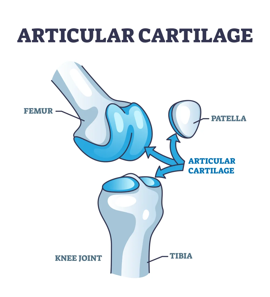
Articular cartilage is the smooth, glassy surface covering the ends of the bones in your knee. It lets the joint glide without friction and absorbs a fair amount of shock with every step. When it’s damaged, doctors describe the injury using a grading scale from I to IV. A grade I injury is a soft spot in otherwise intact cartilage. Grade II involves minor surface tears. Grade III means deeper crevices that don’t yet reach bone. Grade IV, sometimes called a full-thickness lesion, goes all the way down to the bone underneath.
Here’s the part that surprises most patients: cartilage damage often doesn’t hurt at first. Cartilage itself has no nerve supply, so a lesion can sit there quietly for months before it starts causing pain, usually once the exposed bone underneath becomes irritated or a fragment breaks loose and starts floating around the joint.
What I See in My Patients
Several groups walk through my door with this problem. The first are younger, active patients, often athletes here in the Richmond area, who’ve had a specific twisting injury or a fall that damaged a focal area of cartilage. The second are patients in their forties and fifties whose cartilage has simply worn down over years of use, sometimes alongside early arthritis. I treat these groups quite differently, because a 25-year-old athlete with a small, isolated defect has very different goals and healing potential than someone in their fifties with more diffuse wear across the joint.
I’m also careful not to jump straight to imaging severity. I’ve seen MRIs that look alarming attached to patients with barely any pain, and I’ve seen fairly modest-looking lesions in patients who are miserable. The exam and the patient’s actual experience carry as much weight for me as the picture on the screen. Sometimes I find a cartilage lesion during an arthroscopy done for a completely different reason, and in those cases, if it isn’t causing symptoms, I’m generally inclined to leave it alone rather than potentially aggravate something the patient never noticed in the first place.
Where Treatment Starts: Nonsurgical Options
Not every cartilage injury needs surgery right away, even when it’s causing symptoms. I typically start with activity modification, a course of physical therapy aimed at strengthening the muscles around the knee, and sometimes a supportive sleeve or brace. These measures won’t regrow cartilage, but they can meaningfully reduce the load on the damaged area and calm symptoms enough that surgery isn’t necessary, at least for a while. Heat, ice, and over-the-counter anti-inflammatory medication also have a place here, mainly for managing flare-ups rather than fixing the underlying defect.
Reparative Surgery: Cell Stimulation Techniques
When nonsurgical care isn’t enough, the first tier of surgical options aims to stimulate the body’s own healing response, mainly by encouraging fibrocartilage, a tougher and less ideal substitute tissue, to fill the defect.
Arthroscopic debridement simply cleans up rough edges and loose fragments and tends to work best as a shorter-term solution for older patients or larger lesions where a bigger procedure isn’t practical. Microfracture, where I create tiny holes in the bone beneath the lesion with a small awl, triggers a fresh blood supply that helps fibrocartilage form over time. Abrasion arthroplasty works on a similar principle, using a burr to encourage the same healing response. All three are outpatient procedures, and all three tend to provide meaningful, though sometimes temporary, relief.
Restorative Surgery: Rebuilding the Original Cartilage
For younger, more active patients with a well-defined defect, I may lean toward restorative procedures that attempt to replace the missing cartilage with tissue that behaves like the original rather than fibrocartilage.
Osteochondral autografting takes a plug of bone and cartilage from a lower-stress area of your own knee and transplants it directly into the defect in one procedure. For larger defects, particularly cases of osteochondritis dissecans where a chunk of cartilage and the bone beneath it has lost its blood supply, an osteochondral allograft using donor tissue is often the better option. I perform the BioUni procedure for these larger grafts, which allows for a more precise fit than older grafting techniques, and I’ve built a significant amount of experience with allograft surgery over the years.
When the Cartilage Injury Isn’t Acting Alone
Cartilage rarely gets damaged in complete isolation. I frequently find a meniscus tear alongside a cartilage lesion, since the two structures share so much of the same load-bearing role in the knee. In patients further along in years, cartilage wear can be a part of a broader picture of knee osteoarthritis. Part of my job is figuring out which of these pictures I’m actually looking at before recommending a specific procedure.
What Recovery Looks Like
Recovery depends heavily on which procedure we do, but a common thread runs through most of them. Patients typically use crutches and limit weight on the leg during the first part of recovery to protect the healing area, and I often use a continuous passive motion machine early on to keep the joint moving gently and reduce stiffness.
Physical therapy progresses in stages, starting with pain control and protected motion, then building toward strength and, eventually, a return to normal activity. Allograft patients tend to have the longest protected weight-bearing period, generally around six weeks, given the size of tissue involved.
Summary
Knee cartilage damage covers a wide range of severity, from a small soft spot that responds to physical therapy to a full-thickness defect that needs restorative surgery or a graft. The right treatment depends on the grade of the injury, your age, and how you want to use your knee going forward, not on a one-size-fits-all protocol. If you’ve been told you have a cartilage lesion, or if you’re dealing with knee pain that hasn’t responded to rest, the next step is an exam and imaging so we can evaluate the injury accurately. Call my Midlothian office at (804) 939-6651 or schedule an appointment online.
Frequently Asked Questions
Is knee cartilage damage the same thing as a meniscus tear?
No. The meniscus is a separate cushioning structure between the bones, while articular cartilage is the smooth surface coating the ends of the bones themselves. They’re commonly injured together, but they’re treated differently.
Can damaged knee cartilage heal on its own?
Generally not fully. Cartilage has no direct blood supply, so it lacks the healing mechanisms most other tissues rely on. Some smaller injuries can be managed successfully without surgery, but the tissue itself typically won’t regenerate without help.
How do you decide which cartilage procedure is right for a patient?
I weigh the size and grade of the lesion, your age, activity level, and the overall condition of the rest of your knee. A small, isolated defect in an active 30-year-old calls for a different approach than diffuse wear in a 55-year-old with early arthritis.
How long does recovery take after cartilage surgery?
Most patients use crutches and limit weight-bearing for up to six weeks, followed by a structured physical therapy program. Full recovery and return to higher-impact activity generally takes several months, depending on the procedure.