

A knee injury can be a frightening and confusing experience. You might feel pain, notice swelling, and wonder what exactly happened inside the joint. One of the most common knee injuries involves a ligament on the inner side of your knee. This raises an important question for many patients: What is an MCL tear? The medial collateral ligament, or MCL, is a key structure for knee stability, and an injury to it can interfere with your daily life and athletic activities. In this post, we’ll explore the anatomy of the knee, explain how MCL injuries can occur, and detail the symptoms you might experience.
Key Takeaways
- An MCL tear is an injury to the medial collateral ligament, a band of tissue on the inner side of your knee that connects the thighbone to the shinbone and provides stability.
- These injuries are often caused by a direct blow to the outside of the knee or by sudden twisting or cutting motions, common in sports like football, soccer, and skiing.
- Symptoms typically include a popping sound at the time of injury, pain and tenderness on the inner knee, swelling, and a feeling of instability or the knee “giving out.”
- Many MCL tears can heal without surgery due to the ligament’s good blood supply. Treatment often involves rest, ice, bracing, and physical therapy. Surgery may be considered for severe tears.
Understanding the Knee’s Anatomy
To fully grasp what an MCL tear is, it helps to first understand the structure of the knee. The knee is one of the largest and most complex joints in the body. It joins the thighbone (femur) to the shinbone (tibia). The fibula and the patella are the other bones that make up the knee joint.
Within this structure, ligaments act like strong ropes to connect the bones and provide stability. The knee has four primary ligaments:
- Anterior Cruciate Ligament (ACL): Located in the center of the knee, it controls the forward movement and rotation of the shinbone.
- Posterior Cruciate Ligament (PCL): Also in the center of the knee, it controls the backward movement of the shinbone.
- Lateral Collateral Ligament (LCL): This ligament runs along the outer side of the knee.
- Medial Collateral Ligament (MCL): This is the ligament that runs along the inner side of your knee. It is a broad, flat band of tissue that connects the femur to the tibia. Its primary job is to resist force from the outer side of the knee, an action known as valgus force, which prevents the knee from buckling inward.
What is an MCL Tear? The Injury Explained
An MCL tear occurs when this ligament is stretched or torn. Because of its role in stabilizing the inner knee, the MCL is vulnerable to injury when a strong force pushes the knee sideways. The damage can range from microscopic tears in the ligament fibers to a complete rupture of the ligament.
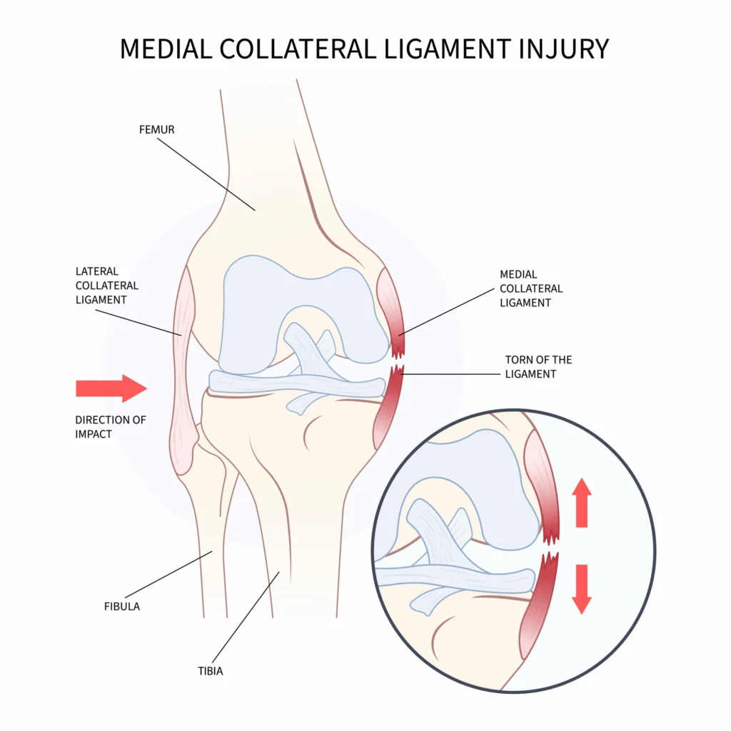
MCL tears may be classified into three grades based on their severity:
- Grade 1 Tear: This is the mildest form of injury. The MCL is stretched, but not significantly torn. There may be tenderness and minor pain on the inner side of the knee, but the joint usually remains stable.
- Grade 2 Tear: This is a moderate injury, involving a partial tear of the MCL. You will likely experience more intense pain and tenderness. The knee joint may feel loose or unstable, especially when pressure is applied.
- Grade 3 Tear: This is the most severe type of MCL injury, indicating a complete tear of the ligament. The knee joint is often very unstable, and you may feel it giving way. Pain can be intense, though sometimes it may decrease after the initial injury because the ligament is no longer under tension.
Common Causes of an MCL Tear
MCL tears can be the result of a sudden, forceful event. The most common cause is a direct blow to the outside of the knee. This impact forces the knee to bend inward, placing extreme stress on the MCL. This scenario is frequently seen in contact sports.
Other common causes include:
- Sudden Twisting or Cutting: Athletes who plant their foot and quickly change direction can put significant strain on the MCL. This is common in sports like soccer, basketball, and football.
- Hyperextension: In sports like skiing, the knee can be forced to bend backward too far, which can lead to an MCL injury.
- Awkward Landings: Landing improperly from a jump can create a valgus force on the knee, leading to a tear.
- Repetitive Stress: While less common, repeated pressure on the knee can strain the ligament, making it more susceptible to injury from minor incidents.
Recognizing the Symptoms of an MCL Tear
The symptoms of an MCL tear can vary depending on the severity of the injury. However, there are several common signs that may indicate you have damaged this ligament.
- A Popping Sound: Some individuals report hearing or feeling a “pop” at the moment of injury.
- Pain and Tenderness: You will likely feel pain directly over the inner side of your knee. The area may be tender to the touch.
- Swelling: The knee may swell soon after the injury.
- Instability: A common sensation is that the knee feels unstable, as if it might “give out” or buckle sideways.
- Locking or Catching: You might feel the knee joint lock or catch as you move it.
- Difficulty Walking: Depending on the grade of the tear, walking can be difficult and painful. With a severe tear, you may not be able to bear weight on the injured leg.
How an MCL Tear is Diagnosed
An accurate diagnosis is important for developing an effective treatment plan. Your doctor will typically begin by discussing your symptoms and how the injury occurred. This will be followed by a physical examination of your knee.
During the exam, your doctor will carefully check for pain and tenderness along the MCL. They may also perform a valgus stress test. In this test, your doctor will apply gentle pressure to the outside of your knee while it is slightly bent. This action tests the stability of the MCL. If the ligament is torn, the inner side of the knee may open up more than it would on an uninjured knee.
In addition to the physical exam, imaging tests may be used to confirm the diagnosis and check for other injuries:
- X-rays: While X-rays do not show ligaments, they may be used to rule out a fracture in the bones of the knee.
- Magnetic Resonance Imaging (MRI): An MRI is an imaging tool for visualizing soft tissues like ligaments. It can show the extent of the MCL tear and identify any associated injuries to the ACL, meniscus, or cartilage.
- Ultrasound: In some cases, an ultrasound can be used to assess the MCL and surrounding structures.
Treatment Options for an MCL Tear
The MCL has a robust blood supply, which gives it a strong potential to heal on its own. As a result, many tears can be treated effectively without surgery. The primary goals of treatment are to reduce pain and swelling, protect the ligament from further injury, and restore knee function.
Nonsurgical Treatment
For most patients, a conservative treatment approach is recommended. This typically involves:
- The RICE Method:
- Rest: Avoid activities that cause pain and limit weight-bearing on the injured knee. Crutches may be recommended.
- Ice: Apply ice packs to the inner knee for 15-20 minutes every few hours to help reduce swelling and pain.
- Compression: Use an elastic bandage to wrap the knee, which can help control swelling.
- Elevation: Keep your knee elevated above the level of your heart as much as possible.
- Bracing: A knee brace is commonly used to protect the healing ligament. For mild sprains, a simple sleeve may suffice. For more severe tears, a hinged brace that limits side-to-side motion while allowing controlled bending and straightening may be prescribed.
- Medication: Over-the-counter nonsteroidal anti-inflammatory drugs (NSAIDs), such as ibuprofen or naproxen, can help manage pain and reduce inflammation.
- Physical Therapy: A physical therapist can guide you through exercises designed to restore your knee’s range of motion, strength, and stability. This may include exercises to strengthen the quadriceps, hamstrings, and hip muscles, all of which support the knee joint.
Surgical Treatment
Surgery for an MCL tear may be considered in specific situations, particularly when a Grade 3 tear is accompanied by other significant knee injuries, such as a torn ACL. If the knee remains unstable despite a course of nonsurgical treatment, surgery might also be an option.
Rehabilitation and Recovery
Recovery from an MCL tear depends on the severity of the injury and the treatment approach. The timeline can vary significantly from person to person.
The rehabilitation process is a progressive journey that gradually reintroduces stress to the healing ligament. Your physical therapy program will likely advance through several phases, from gentle range-of-motion exercises to more advanced strengthening and sport-specific drills. It is important to follow the guidance of your doctor and physical therapist and not rush the process. Returning to activities too soon can risk re-injury.
Preventing MCL Tears
While not all injuries are preventable, there are steps you can take to reduce your risk of an MCL tear, especially if you participate in high-risk sports.
- Strengthening: A well-conditioned leg provides better support for the knee. Focus on strengthening the muscles of the hips, quadriceps, and hamstrings.
- Flexibility: Regular stretching can help maintain good flexibility in the muscles surrounding the knee.
- Proper Technique: In sports, using proper technique for cutting, pivoting, and landing can reduce abnormal stress on the knee.
- Warm-ups and Cool-downs: Always warm up before exercise and cool down afterward to prepare your muscles for activity and aid in recovery.
- Appropriate Footwear: Wear shoes that are appropriate for your sport and provide good support.
Summary
An MCL tear is a common yet significant knee injury that involves damage to the ligament on the inner side of the knee. These injuries can range from a mild sprain to a complete rupture and are often the result of a direct blow or a sudden twisting motion. While the symptoms of pain, swelling, and instability can be alarming, the good news is that the MCL has a strong capacity for healing. Most patients can recover with nonsurgical treatments like rest, bracing, and a dedicated physical therapy program. By understanding the nature of your injury and working closely with an experienced knee specialist, you can navigate the path to recovery.
Frequently Asked Questions
Can I still walk with a torn MCL?
Whether you can walk may depend on the severity of the tear. With a mild Grade 1 tear, you may be able to walk with some pain. A moderate Grade 2 tear can make walking difficult due to pain and instability. With a severe Grade 3 tear, walking is often very difficult and painful, and you may require crutches to avoid putting weight on the leg.
Do all MCL tears require surgery?
No, the vast majority of MCL tears do not require surgery. The MCL has a good blood supply, which allows it to heal effectively with nonsurgical treatments like bracing and physical therapy. Surgery is typically reserved for severe Grade 3 tears that are part of a more complex injury involving other knee ligaments, or for cases where the knee remains unstable after conservative treatment.
What is the difference between an MCL tear and an ACL tear?
The MCL is located on the inner side of the knee, while the ACL is located in the center of the joint. The MCL primarily prevents the knee from buckling inward, whereas the ACL controls forward movement and rotation of the shinbone. While both can be injured in similar ways, the treatment and recovery processes can differ significantly.